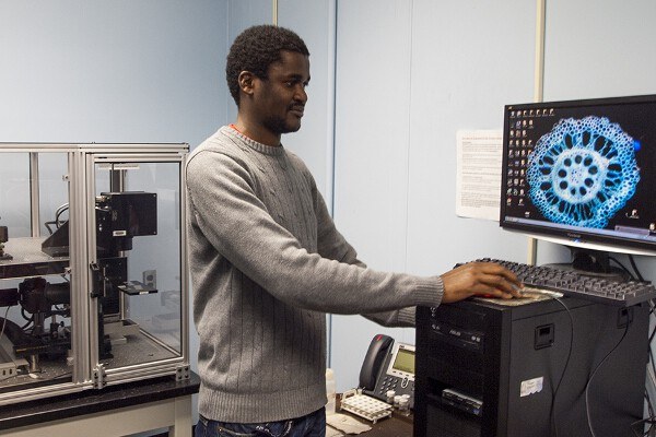
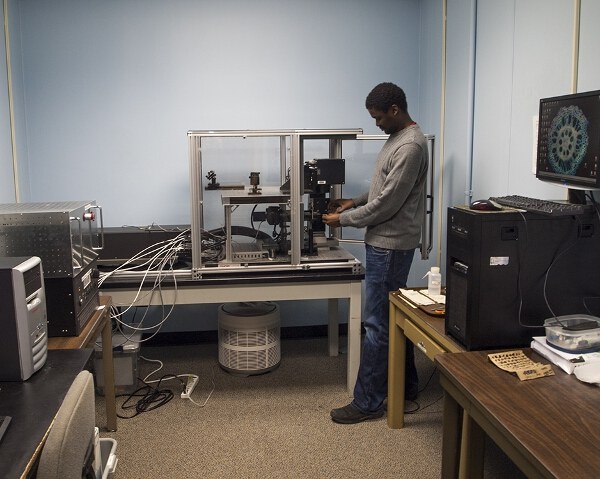
This facility is revolutionizing high-throughput phenotyping of root anatomical traits by increasing the speed of sectioning and imaging and allowing 3D reconstructions.

Using RootScan
An Avia 7000 class 4 355 nm pulsed laser is used to ablate the surface of the cut end of the root sample, revealing a clean, flat surface of the root that is immediately photographed with a Canon DSLR camera and 5x macro lens to capture the entire section. (See more about our laser ablation tomography system in the Methodology section). The resulting digital image is analyzed for anatomical traits using RootScan, a semi-automated image analysis program specifically designed for this purpose (Burton et al., 2012). RootScan currently detects variables such as areas of the cross section, cortex, stele, counts of xylem vessels and aerenchyma lacunae, and cell sizes and counts in radial bands of the cortex as determined by the user.
Maize Root Anatomy Laser Ablation Tomography
Video from Benjamin Hall


